The human body is extremely complex. There are interactions between millions of elements to ensure that every structure works properly and in coordination with the rest of the body. Of course, the brain is no exception; it is, instead, the ideal example of these interactions. The functioning of the brain at the molecule level, from its development through adult life, is not perfectly understood yet. Thus, the discipline of neuroscience is investing great efforts into unraveling every molecular route that leads to the structure of the brain as we know it.
Blood vessels are not only vessels
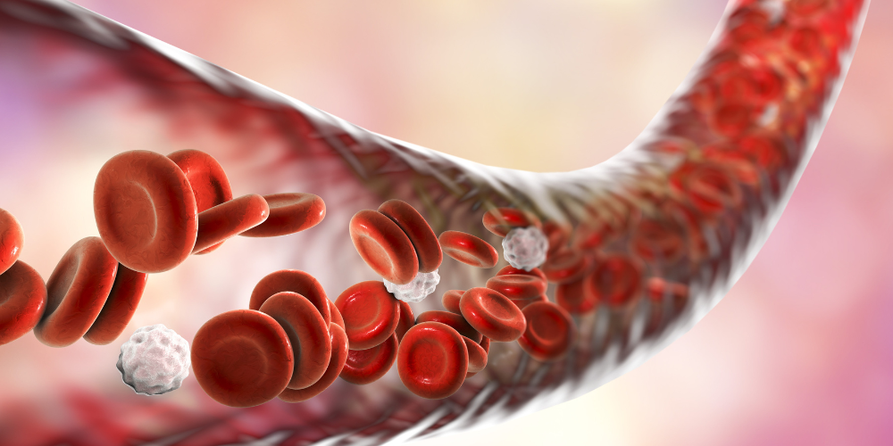
The brain is composed not only by neurons but by many other types of cells that provide support and nutrients, among other functions. As we investigate, we discover more and more functions of these other cell types. A recent discovery showed something surprising: the cells of the vascular system (i.e. the cells of blood vessels) that circulate through the brain tissue have more functions other than constituting the nutrient supply and generating a barrier between blood and brain. It was suggested that the cells of the vascular system could also communicate with neural circuits and regulate them. Thus, vascular cells could directly affect cognition. The phenomenon was discovered in the peripheral nervous system (i.e. the nerves around the body); the next question was, inevitably, if this regulation happened also in the central nervous system (i.e. encephalon, where the brain is the bigger structure, and spinal cord).
Sema 3G, the protein segregated by blood vessel walls
To determine if the regulation of neural circuits by vascular cells happens in the brain, Tan and colleagues conducted the present study. The team selected a protein that is generated only by endothelial cells (i.e. cells of the walls of blood vessels), and not by any other brain cells. The selected protein was Semphorine 3G (Sema 3G), which executes its functions via another protein, neuropilin-2 (Nrp2). Sema 3G is present in the adult brain, for which it may impact the connections between neurons (synapses) in adults.
To study the function of Sema 3G in the brain, the team generated a group of mice lacking the gene responsible for producing the protein. The process is called knockout of Sema 3G.
Once they had the knockout mice, the first thing they did was determining if the animals presented changes in vascular structure and in the blood-brain barrier. After the analyses, no obvious changes were observed in neither of them. Thus, without the protein, the structural aspect of brain blood vessels is not compromised.
VideoTrack as a useful tool for studying Sema 3G in mice
Mice knockout for Sema 3G and normal mice, those without the mutation, acting as controls, were put under behavioral analysis. They did several tests: the Y-maze task, a maze composed of three identical arms where mice were allowed to move freely (they recorded the sequence of arm entries to define alternation); the rotarod test, which examines motor coordination; the pole test, where the mouse is placed on the top of a vertical pole and the time before descending recorded; the tail suspension test, where the mouse is suspended by the tail and its behavior analyzed, with no movement being considered as an indicator for depression; the open field test, where they were placed in the center of a chamber and their movements recorded, allowing for the assessment of locomotor activity and anxiety level; and, finally, the learning and memory test. VideoTrack v3 program was used to automatize the evaluation of most of the mentioned tests. The use of this software ensures unbiased results, speeds up the research process and automatically calculates parameters for each animal. Moreover, it does not use sensors on the animals, for which it does not increase their stress levels. It is the perfect tool for animal behavior analysis.

Sema 3G is necessary for healthy neural connections
The team found, after analysis of the behavioral tests, that hippocampus-dependent memory retrieval (i.e. the retrieval of contextual memories dependent on a brain structure called hippocampus) was significantly impaired in the knockout mice compared to the control ones. The observation led to the following conclusion: Sema 3G is required for normal hippocampal function in the neural connections of adult mice.
On top of the impairment in memory function, they found many other markers that indicated a reduced connectivity between the neurons in the hippocampus; they could observe structural and transmission changes in those neurons. Thus, they concluded that Sema 3G is necessary for neuronal function.
The team also evaluated how Sema 3G produces its effect on hippocampal neurons. They stated that Sema 3G binds to the receptor Nrp2/PlexinA4 in those neurons, which then activates the protein Rac1. Knowing the exact molecular route is important to be able to act on it and modify it.
In sum, the discoveries lead to the conclusion that Sema 3G is essential for normal hippocampal connectivity and hippocampal-related learning and memory. Neurovascular interactions ensure proper neuronal function during development and adulthood.
The future of Sema 3G
Why is this discovery important? The changes observed in knockout mice are very similar to the abnormalities found in various neurodegenerative and psychiatric disorders. For instance, the abnormalities could account for the cognitive decline and memory loss observed in Alzheimer's disease or schizophrenia. This finding provides clinical insight into a potential role for Sema 3G in cognitive dysfunction, and opens the door to Sema 3G as a future therapeutic target. Future studies should research if stimulation of Sema 3G has clinical benefits for maintaining normal neuronal connectivity and normal cognitive behavior.
References
Tan C, Lu N, Wang C, Chen D, Sun N, Lyu H, Körbelin J, et al. (2019). Endothelium-Derived Semaphorin 3G Regulates Hippocampal Synaptic Structure and Plasticity via Neuropilin-2/PlexinA4. Neuron.101(5); 920-937. doi: 10.1016/j.neuron.2018.12.036


