New evidence indicates cacna1aa loss-of-function zebrafish is an ideal animal model of absence seizures and proves beneficial for in vivo drug screening. For the first time, researchers have demonstrated the behavioural and phenotypic alterations of reduced cacna1aa larval zebrafish.
New evidence indicates cacna1aa loss-of-function zebrafish is an ideal animal model of absence epilepsy and proves beneficial for in vivo drug screening. For the first time, researchers have demonstrated the behavioural and phenotypic alterations of reduced cacna1aa larval zebrafish.
What is cacna1a ?
The gene cacna1a encodes the a1 subunit of the voltage gates P/Q type Ca2+ channels (Heyes et al., 2015. These Ca2+ channels are abundant in the presynaptic terminals, particularly in cerebellum's Purkinje cells where they regulate transmitter release (Hillman et al., 1991; Takahashi and Momiyama, 1993; Volsen et al., 1995, Westenbroek et al., 1995). Studies have reported cacna1a mutations in patients with autosomal-dominant diseases such as spinocerebellar ataxia type 6, episodic ataxia type 2, and familial hemiplegic migraine type 1 (Jen and Wan, 2018).
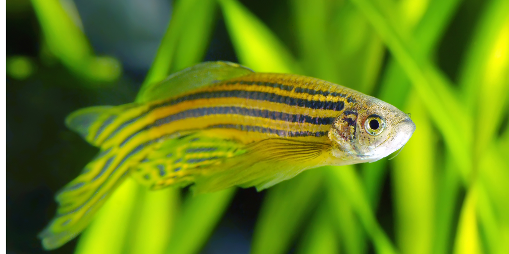
Why zebrafish is an emerging, attractive and robust animal model of absence seizures?
Emerging research has considered zebrafish ideal for modelling human brain disorders. The zebrafish genome is completely sequenced and annotated and shows approximately 70% resemblance with the human genome. Concerning the develpmental stages in zebrafish, 3-day after fertilization corresponds to birth in humans and thereafter every successice day corresponds to 3 monts of human age. For example, 4 and 5 days after fertilizationin zebrafish correspond to 3 and 6 months of age in human children.
Although absence epilepsy has good rodent models, spike-wave discharges (a typical pattern observed on electroencephalograms during an epileptic seizure) in these models appear at the age of 2–3 months, which correspond to juvenile developmental stages in humans. This is relatively late as absence epilepsy manifests itself during early childhood in humans. The researchers in this study took advantage of the opportunity and developed a novel zebrafish model of absence epilepsy that models its characteristics in early larval zebrafish.
Cacna1aa expression in the zebrafish brain
During evolution, the cacna1a gene in zebrafish was duplicated into cacna1aa (72.01%) and cacna1ab (71.28%), homology with the human cacna1a gene. As there is a lack of data for cacna1aa expression in zebrafish, the researchers in this study focused their attention on cacna1aa expression at experimental levels i.e. 4 and 5 days post-fertilization (dpf). They observed noticeable cacna1aa mRNA expression in the hindbrain and midbrain but low in the retina and forebrain. Additionally, they observed high cacna1aa mRNA expression levels in the optic tectum with even higher levels in the medulla oblongata.
Combining two antisense morpholino oligomers (MO), the researchers targeted the start codon (ATG) of all splice variants to achieve partial cacna1aa knockdown (morphants). They then assessed whether this partial loss triggers an epileptic-like phenotype at both the EEG and behavioural levels in larval zebrafish. To further evaluate epileptiform-like discharges, they even studied the activity of four recommended and one contraindicated antiseizure drugs (ASDs) used for treating human absence seizures.
Mortality assessment and touch response of the morphants
To identify an appropriate MO, the researchers administered different concentrations of MO and assessed the mortality rate and touch response of the morphants. Administration of 7.5 ng MO1 did not affect the embryo and larval mortality rate, but at a higher dose of 9 ng the mortality rate enhanced significantly. The concentrations of 7.5 ng and 9 ng hampered touch response in the larval zebrafish. When the researchers introduced another MO, MO2, it did not affect the mortality at both 7.5 and 9 ng, but when raised to 12 ng, they observed a slight increase in mortality. The touch response observed was similar to the hampered effect at 9 ng and 12 ng concentrations.
Further, they combined MO1 and MO2 at doses of 2.5 ng + 2.5 ng, which did not increase the mortality. However, the researchers observed a significant rise (60%) in mortality when they increased the combined dose to 4.5 ng + 4.5 ng after 24 hours post-fertilization. The combined dose of 2.5 ng did not affect the larval touch response and therefore they chose this combination for further experimentation.
Morphological assessment of cacna1aa morphants
The cacna1aa morphants with single MO doses of 9 or 12 ng showed significant morphological malformations such as small heads, curved body axis, yolk sac malformation, pericardial edema and tiny eyes.
Alternatively, morphants with combined MO dose (2.5 ng + 2.5 ng) were slightly hyperpigmented compared to the control ones. Moreover, most morphants did not inflate their swim bladder and had a shorter body length.
ZebraBox assesses behavioural characteristics of cacna1aa morphants.
Zebrabox investigated the locomotor activity of both morphants as well as the control group. Zebrabox is a high-throughput monitoring system that studies zebrafish behaviour. The researchers recorded the distance covered by each larva in two phases; 5 min light phase and 5 min dark phase.
Studies have demonstrated that sudden switching from the light phase to dark phase induces a rapid increase in locomotor activity. The researchers observed that the control group exhibited increased activity after switching from light to dark phase, but cacna1aa morphants demonstrated a decrease in activity compared to the control group. Moreover, the morphants did not show any increase in activity in response to sudden switching from light to dark phase.
EEG discharges and effects of ASDs on cacna1aa morphants
Epileptiform-like events such as spikes and sharp waves are hallmark EEG characteristics of a seizure focus. The researchers recorded involuntary epileptiform-like events such as spike-wave complexes, polyspike-wave discharges and high-voltage spikes in 92% of cacna1aa morphants.
Next, to evaluate its efficiency as a novel animal model for drug screening, the researchers studied the activity of four effective ASDs ( ethosuximide, topiramate, lamotrigine and sodium valproate) that are recommended for absence seizures and one contraindicated drug for the observed type of seizure.
All four effective ASDs significantly decreased the frequency of epileptiform-like discharges, and as expected carba mazepine did not alleviate any seizure parameters.
Could partial cacna1aa knockdown zebrafish be a novel drug screening animal model for absence seizures?
Although the medical community considers absence seizures as a mild form of epilepsy, it is often associated with persistent comorbidities. Moreover, drugs for treating absence seizures such as VPA and ETX are ineffective or not well tolerated. Therefore, this zebrafish model is a promising candidate to imitate absence seizures observed in children with possible applications in evaluating associated comorbidities such as anxiety and memory and learning deficits.
References
1. Gawel, K., Turski, W.A., van der Ent, W., Mathai, B.J., Kirstein-Smardzewska, K.J., Simonsen, A., Esguerra, C.V., 2020. Phenotypic Characterization of Larval Zebrafish (Danio rerio) with Partial Knockdown of the cacna1a Gene. Mol. Neurobiol. 57, 1904–1916. https://doi.org/10.1007/s12035-019-01860-x 2. Heyes, S., Pratt, W.S., Rees, E., Dahimene, S., Ferron, L., Owen, M.J., Dolphin, A.C., 2015. Genetic disruption of voltage-gated calcium channels in psychiatric and neurological disorders. Prog. Neurobiol. 134, 36–54. https://doi.org/10.1016/j.pneurobio.2015.09.002 3. Hillman, D., Chen, S., Aung, T.T., Cherksey, B., Sugimori, M., Llinás, R.R., 1991. Localization of P-type calcium channels in the central nervous system. Proc. Natl. Acad. Sci. 88, 7076–7080. https://doi.org/10.1073/pnas.88.16.7076 4. Jen, J.C., Wan, J., 2018. Chapter 13 - Episodic ataxias, in: Manto, M., Huisman, T.A.G.M. (Eds.), Handbook of Clinical Neurology, The Cerebellum: Disorders and Treatment. Elsevier, pp. 205–215. https://doi.org/10.1016/B978-0-444-64189-2.00013-5 5. Takahashi, T., Momiyama, A., 1993. Different types of calcium channels mediate central synaptic transmission. Nature 366, 156–158. https://doi.org/10.1038/366156a0 6. Volsen, S.G., Day, N.C., McCormack, A.L., Smith, W., Craig, P.J., Beattie, R., Ince, P.G., Shaw, P.J., Ellis, S.B., Gillespie, A., Harpold, M.M., Lodge, D., 1995. The expression of neuronal voltage-dependent calcium channels in human cerebellum. Mol. Brain Res. 34, 271–282. https://doi.org/10.1016/0169-328X(95)00234-J 7. Westenbroek, R., Sakurai, T., Elliott, E., Hell, J., Starr, T., Snutch, T., Catterall, W., 1995. Immunochemical identification and subcellular distribution of the alpha 1A subunits of brain calcium channels. J. Neurosci. 15, 6403–6418.